Ultrasound Imaging: How It Works and Its Clinical Importance
What Is Ultrasound Imaging and How Does It Work?
Ultrasound imaging, also known as sonography, utilizes high-frequency sound waves to create images of the inside of the body. This technique is non-invasive, meaning it doesn't require any incisions or needles, making it a patient-friendly option. The ultrasound machine sends sound waves through a gel applied to the skin, which then reflects back to create a detailed image of organs and tissues.
Ultrasound imaging is a powerful tool that allows us to visualize the human body in real-time, providing critical information for diagnosis and treatment.
These sound waves travel at different speeds depending on the type of tissue they encounter. For instance, they travel faster through bone and slower through fluid. The differences in these speeds are what allow the ultrasound machine to construct an accurate image, much like how a bat uses echolocation to navigate in the dark.
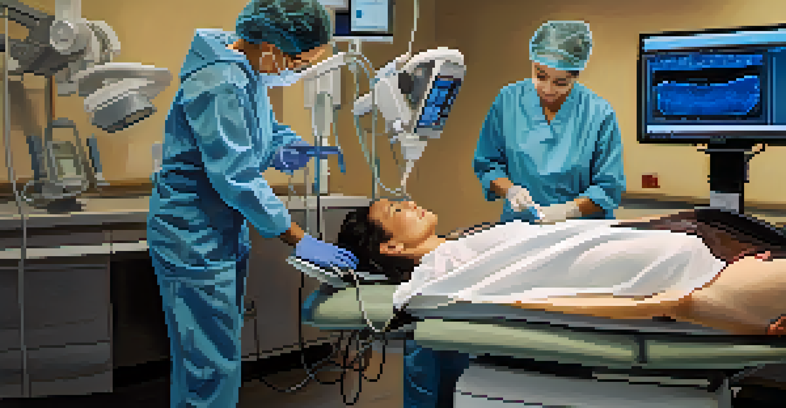
With the help of a trained technician, the images produced can be viewed in real-time on a monitor, providing immediate insights that can guide further medical decisions. This technology is widely used in various medical fields, including obstetrics, cardiology, and emergency medicine.
The Equipment Used in Ultrasound Imaging
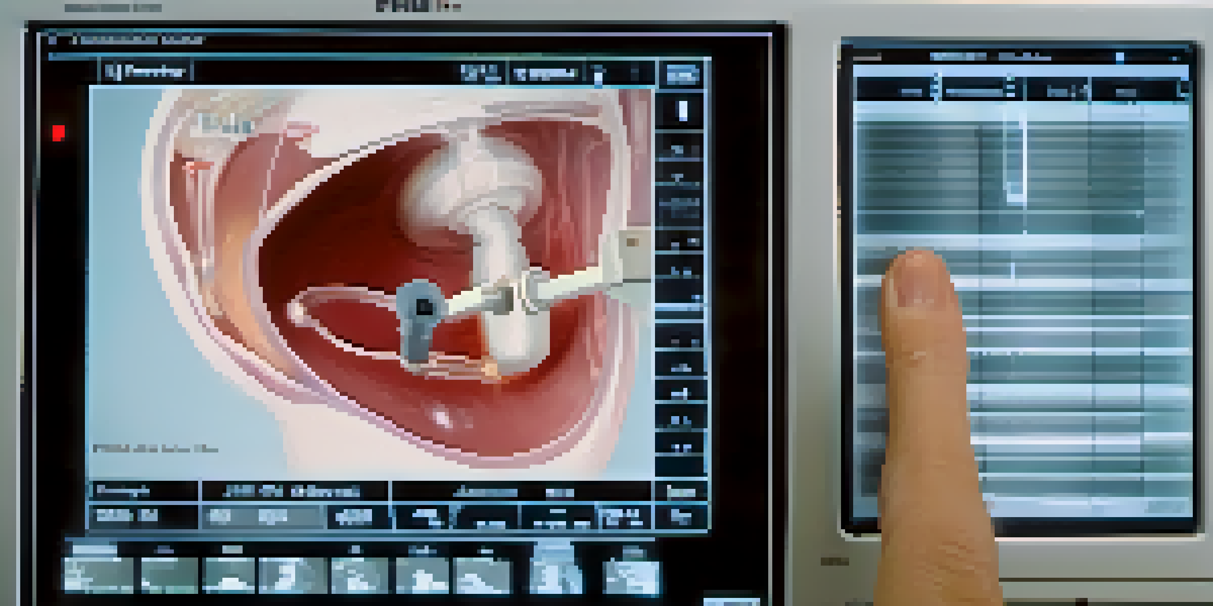
An ultrasound machine consists of several key components: a transducer, a computer, and a display monitor. The transducer is a handheld device that emits sound waves and receives the echoes that bounce back from the body's tissues. It’s akin to a microphone that both sends out signals and listens for responses, ensuring a clear picture is formed.
The computer processes the received echoes and converts them into visual images. This processing is crucial because it translates the raw sound data into a format that healthcare providers can interpret easily. Think of it as translating a foreign language into something you can understand.
Ultrasound: Safe and Non-invasive
Ultrasound imaging uses high-frequency sound waves to create detailed images without ionizing radiation, making it a patient-friendly option.
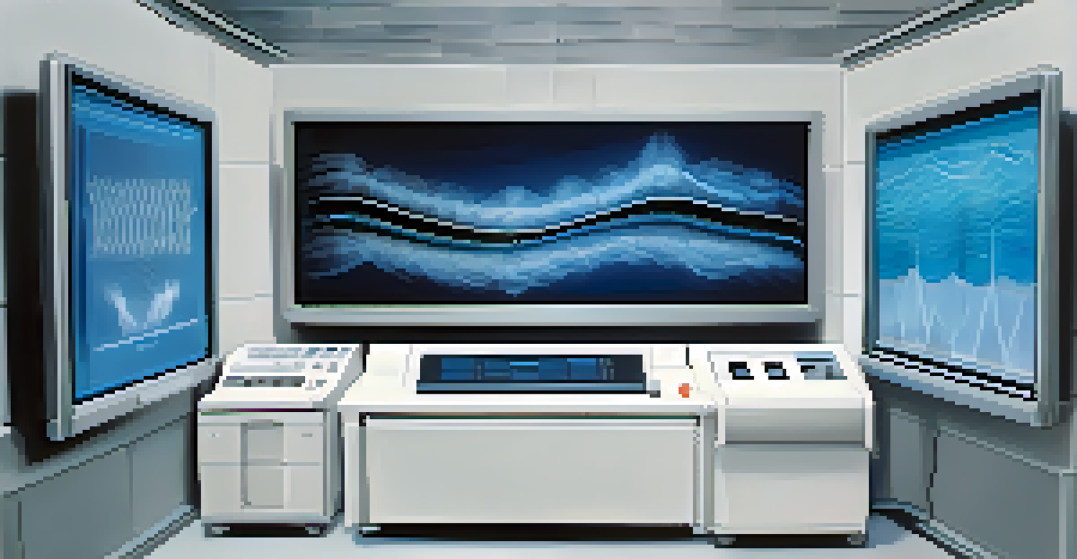
Finally, the display monitor presents these images in real-time, allowing healthcare professionals to analyze the results on the spot. This immediate feedback can be critical for making timely diagnoses and treatment decisions.
Types of Ultrasound Imaging Techniques
There are several types of ultrasound techniques, each tailored for specific diagnostic needs. The most common is the abdominal ultrasound, which helps visualize organs like the liver and kidneys. Additionally, obstetric ultrasound is widely recognized for monitoring fetal development during pregnancy.
The future of ultrasound technology holds great promise, with innovations that will enhance accessibility and improve patient care across various settings.
Another technique, known as Doppler ultrasound, measures blood flow through vessels and is essential for assessing conditions like deep vein thrombosis or heart problems. This method adds a layer of functionality by not only providing images but also illustrating how blood moves through the body.
Transvaginal and transrectal ultrasound techniques are also used for more detailed examinations of reproductive organs. These methods allow for closer and clearer imaging, especially in cases where traditional external ultrasound may not suffice.
Applications of Ultrasound Imaging in Healthcare
Ultrasound imaging has a wide array of applications in healthcare, making it an invaluable tool for diagnosis and treatment. In obstetrics, it provides vital information about fetal health and development, helping expectant parents and healthcare providers monitor pregnancy progress. The ability to visualize the fetus allows for early detection of potential issues.
In addition to obstetrics, ultrasound is frequently used in cardiology to assess heart function. Echocardiograms, a type of cardiac ultrasound, help doctors evaluate the heart's structure and blood flow, guiding treatment decisions for various heart conditions. Think of it as getting a snapshot of your heart’s health in real-time.
Real-time Imaging Enhances Care
The ability to view ultrasound images in real-time allows healthcare professionals to make immediate and informed decisions regarding patient treatment.
Moreover, ultrasound plays a critical role in guiding certain medical procedures, such as biopsies or fluid drainage. By providing a live view of the area being treated, it enhances precision and reduces the risk of complications.
Benefits of Ultrasound Imaging Over Other Methods
One of the primary advantages of ultrasound imaging is that it is safe and non-invasive. Unlike X-rays or CT scans, ultrasound doesn’t use ionizing radiation, which can be harmful in high doses. This makes it an excellent choice for monitoring pregnant women and children, who are more sensitive to radiation.
Additionally, ultrasound is relatively quick and cost-effective compared to other imaging methods. The procedure can often be completed in less than 30 minutes, allowing for faster diagnosis and treatment. This efficiency can be crucial in emergency situations where time is of the essence.
Lastly, the ability to obtain real-time images allows for immediate decision-making. This dynamic aspect of ultrasound imaging can significantly enhance patient care, as healthcare providers can react promptly to emerging issues.
Limitations of Ultrasound Imaging
While ultrasound imaging offers numerous benefits, it does come with certain limitations that healthcare providers must consider. One significant drawback is that ultrasound is highly operator-dependent; the skill and experience of the technician can greatly influence the quality of the images produced. This variability means that results can differ from one provider to another.
Moreover, ultrasound may not provide the same level of detail as other imaging techniques like MRI or CT scans. For instance, while ultrasound is excellent for visualizing soft tissues, it may struggle with certain structures like bones, which can obscure the images. This limitation can sometimes lead to the need for follow-up imaging using different modalities.
Limitations of Ultrasound Imaging
Despite its benefits, ultrasound's effectiveness can be impacted by operator skill and certain physical conditions, which may necessitate further imaging.
Lastly, certain factors, such as obesity or the presence of gas in the intestines, can hinder the effectiveness of ultrasound imaging. These obstacles can result in suboptimal images, making it challenging to obtain a clear understanding of the area being examined.
The Future of Ultrasound Imaging Technology
The future of ultrasound imaging looks promising, thanks to advancements in technology and research. Innovations such as portable ultrasound devices are making it easier for healthcare providers to perform scans in various settings, including remote locations. This accessibility can greatly enhance patient care in underserved areas.
Additionally, improvements in imaging software are enhancing the quality and clarity of ultrasound images. New algorithms and artificial intelligence are being integrated into ultrasound systems to assist in image analysis, potentially increasing diagnostic accuracy. It’s similar to how smartphone cameras have evolved, becoming smarter and more efficient in capturing high-quality images.
Furthermore, researchers are exploring new applications for ultrasound, including therapeutic uses such as targeted drug delivery and tissue regeneration. These developments could redefine how we think about ultrasound in medicine, expanding its role beyond diagnostics to treatment as well.