PET Scans: The Technology Behind Functional Imaging
What is a PET Scan and How Does It Work?
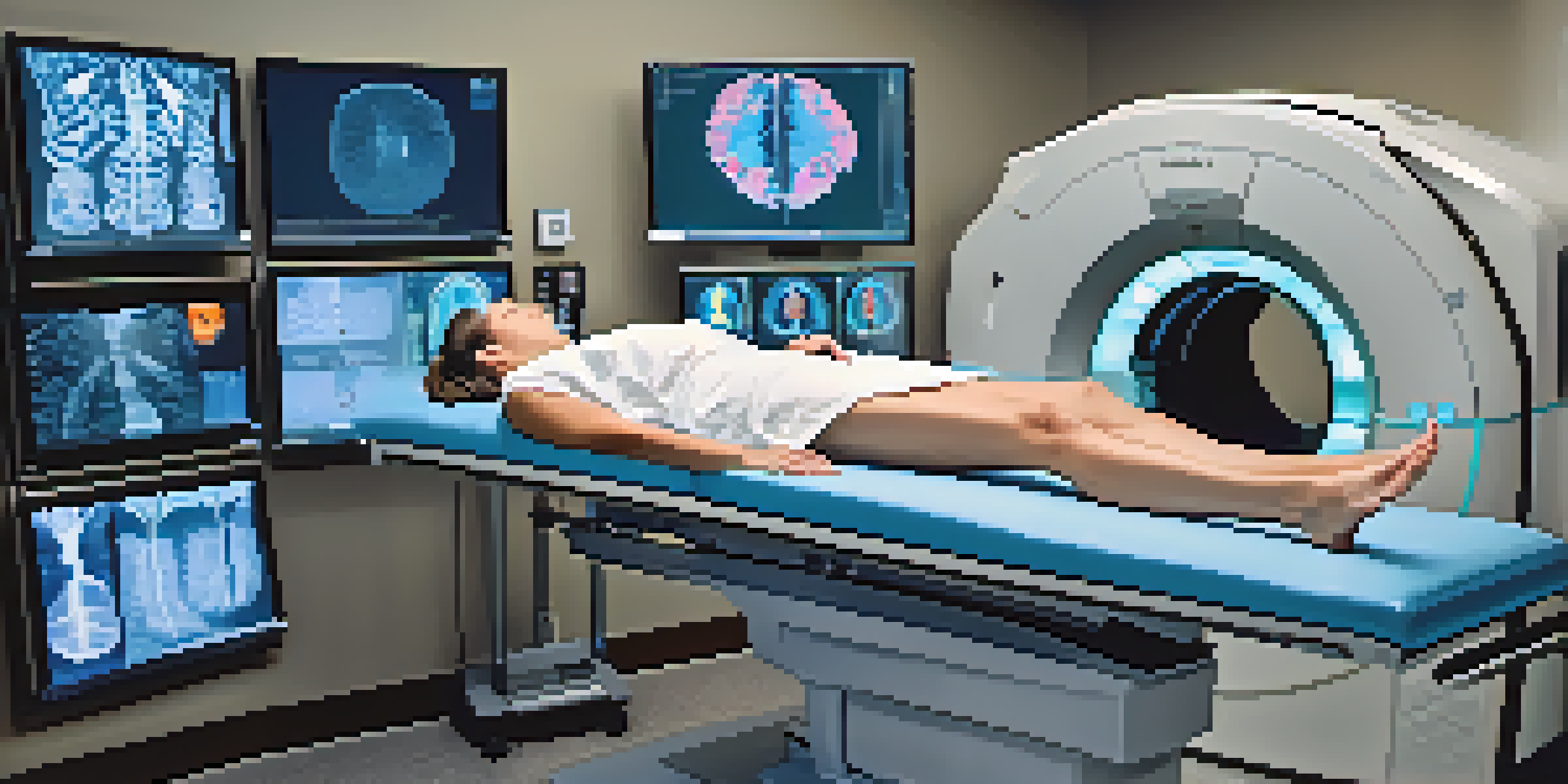
A PET scan, or Positron Emission Tomography scan, is a medical imaging technique that allows doctors to observe metabolic processes in the body. Unlike traditional imaging methods that focus on structure, PET scans provide insights into how tissues and organs function. This is achieved by using a small amount of radioactive material, which emits positrons that are detected by the scanner.
Imaging is a key part of the diagnostic process that can provide insights beyond what we can see with our eyes.
When a patient undergoes a PET scan, they are usually injected with a radiotracer, often a form of glucose. Cancer cells, which have a higher metabolic rate than normal cells, absorb more of this tracer, allowing them to be highlighted during the scan. This makes PET scans particularly useful for detecting cancer, monitoring treatment responses, and evaluating brain disorders.
The images generated from a PET scan are then combined with CT or MRI scans to provide a comprehensive view of the area being examined. This fusion of images enhances diagnostic accuracy, enabling physicians to make informed decisions about treatment options.
The Role of Radiotracers in PET Scans
Radiotracers are the cornerstone of PET scan technology. These substances are designed to highlight specific biological processes in the body, which is essential for accurate diagnosis. The most commonly used radiotracer is fluorodeoxyglucose (FDG), a radioactive form of glucose that cancer cells readily absorb.
The choice of radiotracer can vary based on the condition being investigated. For example, different tracers may be used for brain imaging versus cancer detection. This specificity allows physicians to tailor the imaging process to each patient’s needs, improving the chances of detecting abnormalities.
Understanding PET Scans
PET scans are advanced imaging techniques that reveal how organs and tissues function, particularly useful in detecting cancer and evaluating brain disorders.
By understanding how different radiotracers work and their applications, healthcare providers can better interpret the results of PET scans. This knowledge ultimately leads to more precise treatments and improved patient outcomes.
Key Benefits of PET Scans in Medicine
PET scans offer several advantages over traditional imaging techniques. One significant benefit is their ability to detect diseases at an early stage, often before symptoms arise. This early detection is crucial for conditions like cancer, where timely intervention can significantly improve survival rates.
PET scans have transformed the way we diagnose and manage diseases, giving us a window into the biological processes of the body.
Additionally, PET scans provide functional information about organs and tissues, allowing for a more comprehensive understanding of a patient's health. For instance, in neurology, PET scans can reveal how well the brain is functioning, which is essential for diagnosing conditions like Alzheimer’s disease.
These scans are also non-invasive and generally safe, making them a preferred choice for many diagnostic procedures. As technology advances, the benefits of PET scans continue to expand, offering new possibilities for patient care.
Exploring the Applications of PET Scans
The applications of PET scans are diverse, covering various fields of medicine. In oncology, they are invaluable for detecting tumors, assessing their size, and determining how well treatment is working. This information is crucial for oncologists when deciding on the next steps in a patient's treatment plan.
In addition to cancer, PET scans play an essential role in cardiology. They help evaluate heart function and blood flow, aiding in the diagnosis of conditions such as coronary artery disease. This application showcases the versatility of PET imaging in different medical specialties.
Radiotracers are Essential
Radiotracers, like FDG, are crucial for highlighting metabolic processes in the body, allowing for tailored imaging based on the specific condition being investigated.
Moreover, PET scans are increasingly being used in research settings, contributing to the development of new therapies and understanding of diseases. As researchers explore the intricacies of various conditions, PET imaging provides vital insights that can lead to breakthroughs in treatment.
Understanding the Risks and Limitations of PET Scans
While PET scans are generally safe, there are some risks and limitations to consider. The most notable concern is the exposure to radiation from the radiotracers used in the scans. However, the amount of radiation is typically low and is outweighed by the potential benefits of accurate diagnosis.
Additionally, not all abnormalities can be detected by PET scans. Some conditions may not show up clearly, especially in early stages or if the radiotracer is not absorbed as expected. This limitation emphasizes the importance of using PET scans in conjunction with other diagnostic tools.
Patients should also be aware of potential allergic reactions to the radiotracer, though this is rare. Discussing any concerns with a healthcare provider can help ensure that the benefits of a PET scan outweigh the risks.
The Future of PET Scanning Technology
As technology continues to advance, the future of PET scanning looks promising. Innovations in radiotracer development are paving the way for more specific and effective imaging. Researchers are exploring new compounds that can target various biological processes, enhancing the diagnostic capabilities of PET scans.
Furthermore, improvements in imaging technology are leading to higher resolution images and faster scan times. These advancements not only improve patient comfort but also increase the accuracy of diagnoses, making PET scans an even more valuable tool in medical practice.
Benefits and Limitations
While PET scans offer early disease detection and comprehensive insights, they come with some risks including radiation exposure and limitations in identifying certain abnormalities.
In summary, the evolution of PET scanning technology holds great potential for enhancing patient care. As new techniques and applications emerge, we can expect PET scans to play an increasingly vital role in the future of medicine.
Preparing for a PET Scan: What to Expect
If you’re scheduled for a PET scan, knowing what to expect can help ease any anxiety. Preparation usually involves fasting for several hours before the procedure, as this ensures optimal absorption of the radiotracer. Your healthcare provider will give you specific instructions based on your individual case.
During the scan, you will be positioned comfortably on a table that slides into the PET scanner. It’s essential to remain still during the scan to obtain clear images, which typically lasts around 30 minutes to an hour. Many patients find the experience straightforward and quick, especially with today’s advanced technology.

After the scan, you can usually return to your normal activities immediately. Your healthcare provider will review the images and discuss the results with you during a follow-up appointment. Understanding this process can make your experience smoother and more manageable.